Interview with Peter Atkinson
Q: Can you tell me about yourself, your background and how you became a veterinarian?
After Senior School in Warwick Queensland, I spent two years at Longreach Pastoral College, then a couple of years in northern and Western Queensland working on cattle properties, then another two years working in the family cattle AI and bull semen retailing business. I became very interested in cattle reproduction and decided that a degree in Veterinary Science was the best way of ensuring a long term informed future in the industry. I graduated from the University of Queensland in 1996.
Q: In your practice, you offer a range of reproductive services, can you describe those services?
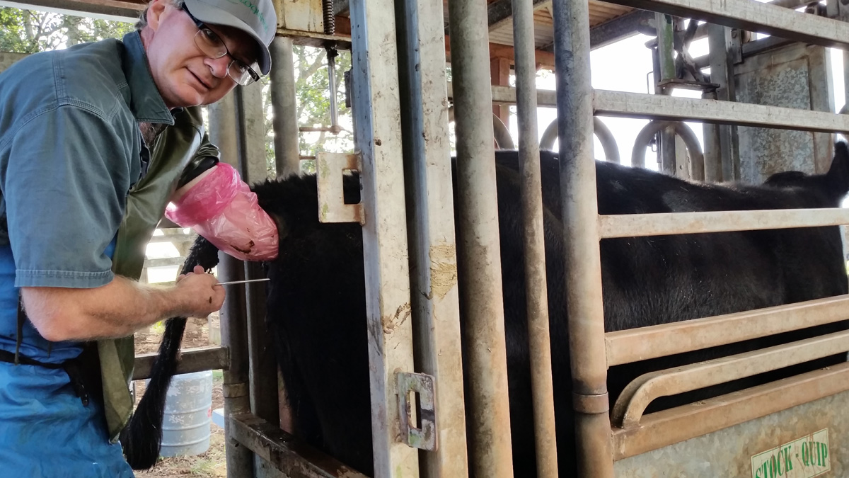
We provide an embryo recovery and transfer service for cattle using a MOET harvesting technique and a non-surgical approach for transfer of embryos into the recipients. We conduct AI programs, pregnancy diagnosis, ultrasound foetal sexing between 8 and 11 weeks. We also provide a bull examination and semen custom collection service.
Q: You have become reliant on ultrasound to provide a high quality and reliable service to your customers. What type of ultrasound do you use in your practice?
We are currently using a Sonosite M Turbo ultrasound machines with a variable 5 -10 MHz linear array probe for our general ET and AI work and a sector probe with an IVF tool for aspirating persistent follicular cysts.
Q: What made you consider starting using ultrasound through procedures that typically would not be used?
We know that skills in trans-rectal palpation of the ovary and uterus are very important, but we found that good ultrasound images of these structures gave us extra information on how to treat donor and recipient cows. Unfortunately, even excellent palpation skills were not sensitive enough to provide the detailed information we needed.
Q: For what procedures do you use ultrasound?
If we are standing at the back of a cow there is almost always an ultrasound on the table beside us. If you don’t look you can’t know!!
Q: How has your understanding of the follicular dynamics improved your management of AI programs – in both heat detection and FTAI programs?
The wave-like nature of follicular development poses some interesting challenges for AI programs. For AI programs we need to provide a single dominant follicle at the time of AI. We are time poor, so do not encourage natural or “prostaglandin only” based programs because of the variation in the onset of standing heats. We therefore use FTAI programs whenever possible. The medications used in fixed time AI programs need to manipulate the ovaries to produce a follicle that is 10 to 14 mm in diameter at the predetermined time of insemination. Unfortunately, not all cows will produce a dominant follicle of this size. If the semen being used is rare of very expensive we can actively select those cows with the most chance of conceiving based on their follicular diameter.
Q: What techniques of ultrasound are you using to interpret follicular and luteal function?
We have been using colour Doppler in an attempt to measure blood flow in the follicular edge at AI and in the CL before embryo transfer if we are unsure about the quality of these structures. The colour doppler technique is interesting but still remains experimental in our hands. Perhaps purchasing an ultrasound with advanced colour doppler functions may improve the speed and repeatability of the technique.
Q: Embryo transfer is a large part of your business, how have you used ultrasound to manage the donor cow?
It enables us to quantify the response to FSH treatment at the time of AI. We assess the size and number of follicles on the donors ovaries at first insemination. At second insemination “12 hours later” we can determine if the follicles are still present on the ovaries or if the follicles have ovulated during the night. In larger flush programs this allows us to flush cows in groups depending on their time of ovulation. It allows us to choose which cows we can flush early on day 6 and not have very young embryos and which cows we flush last on day 7 and not have very old or hatching embryos.
Q: How have your used ultrasound to manage the recipient cow?
Yes, we use the ultrasound on every recipient. Before transferring the embryo into a recipient we will assess presence, size and the central fluid within the CL. If there are CL’s on both ovaries we can select the one with the most luteal tissue. We also check for uterine anomalies such as pus or signs of recent abortion Following up on programs with pregnancy diagnosis, is a relatively mainstream veterinary procedure.
Q: What added benefits do you provide to your clients through this use?
Firstly the clients like to see the foetal images. Very good PR!!! We like to pregnancy test the recipients or AI cows at 11 weeks allowing us to reliably differentiate those pregnancies from the backup bull at 8 and 5 weeks. We can foetal sex with ultrasound between 8 and 11 weeks gestation. We can tell if the foetus is alive “Beating heart” or dead “No beating heart” and if the foetus is degenerating.
Q: How do your other vets in your practice utilise the ultrasound? Was it easy for them to adopt?
We all use the ultrasound machine in the same way. My colleagues adopted the process readily, but it does take some time and patience to become quick and efficient with an ultrasound machine.
Q: Many veterinarians will not use ultrasound due to the fact that it can slow the process down, can you comment?
Yes, I think it will initially make you slower, but I believe the results are better with its use over time. We work in a purely results driven industry and the client will readily forget about the later finish when the conception rates are consistent.
Rate This Content
Register for Updates
