Cattle
Reproductive efficiency in beef and dairy cattle is measured by a cow’s ability to have a calf every 12 months, whilst continuing to achieve her production for beef and/or milk. The higher percentage of females that can achieve this in a herd, the better the reproductive efficiency.
If you plot the reproductive activities of a cow over 12 months (Figure 1), it becomes clear that on average the time frame for conception to achieve this goal is relatively short (55 days). If gestation periods are longer, such as in Bos indicus genotypes, or cows fail to cycle for a long duration after calving, this window becomes even shorter. Breeders must ensure that their cows are managed so that they can conceive within this tight window.
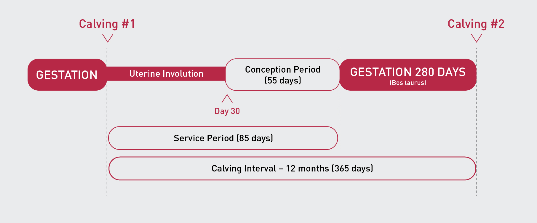
Figure 1: The 12 month reproductive cycle of a productive cow.
A basic understanding of the oestrous cycle and the hormonal interactions can assist in understanding how to manage cattle during this conception period. Alternatively, it can also give reason to using artificial intervention to improve the chances of conception to ensure a cow has a calf on a yearly basis.
Physiology
The oestrous (or oestrus) cycle:
The oestrous cycle in cattle is the period from one oestrous (or heat) to the next one. On average the oestrous cycle of cows/heifers occurs every 21 days, but ranges from 18 to 24 days. There are a series of phases within the oestrous cycle:
-
Luteal phase: commences shortly after ovulation (~ day 1 to 17). After ovulation a Corpus Luteum (CL) forms, secreting progesterone (P4). During this phase the cow/heifer does not exhibit any oestrus behaviour.
-
Follicular phase: commences after the regression of the CL (~ day 18 to 21). The pre-ovulatory follicle emerges, secreting oestradiol (E2), which causes the cow/heifer to demonstrate oestrus behaviour. Near this onset of oestrus, a surge in Luteinising Hormone (LH) occurs. Ovulation usually occurs 24 to 32 hours thereafter.
-
Ovulation: Once the pre-ovulatory follicle has matured and grown to the maximum size, it will ovulate in response to a surge in LH. The oestrous cycle then re-commences.
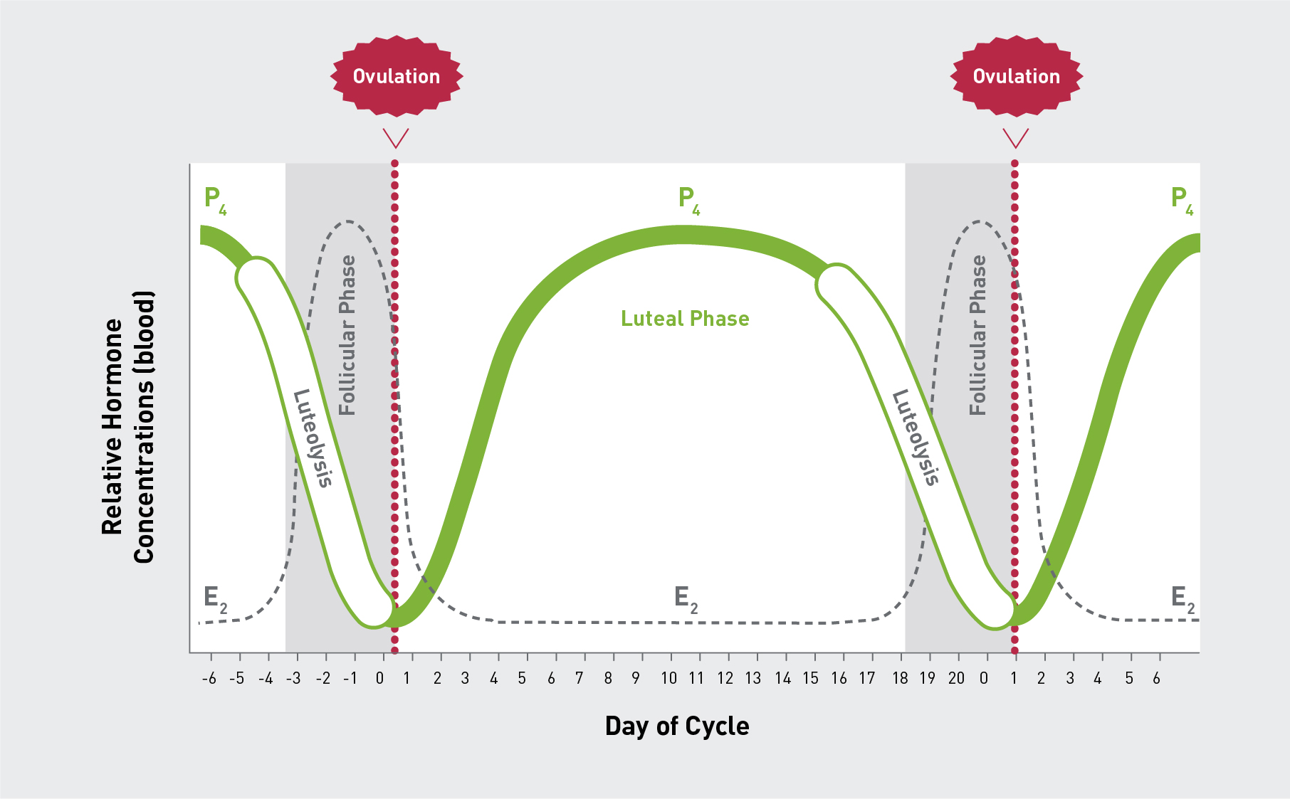
Figure 2: Basic description of the oestrous cycle of the cow
During the oestrous cycle there are usually 2 to 3 follicular waves that occur successively. The first follicular wave commences shortly after ovulation and persists for 8 to 10 days. The follicular wave is initiated by a peak in circulating Follicle Stimulating Hormone (FSH). It is called a follicular wave as a pool of follicles is recruited, from which a single follicle is selected and becomes dominant, whilst the subordinate follicles will become atretic. If there is a CL present at the time this follicle becomes dominant, it will also become atretic, and a new wave will emerge. If there is no CL, the dominant follicle will continue to grow, mature and ovulate. Cattle that have shorter oestrous cycles (i.e. 18 days duration) are likely to have only 2 follicular waves per cycle, whereas cattle with longer oestrous cycles (i.e. 20 days) are likely to have 3 or 4 follicular waves [6].
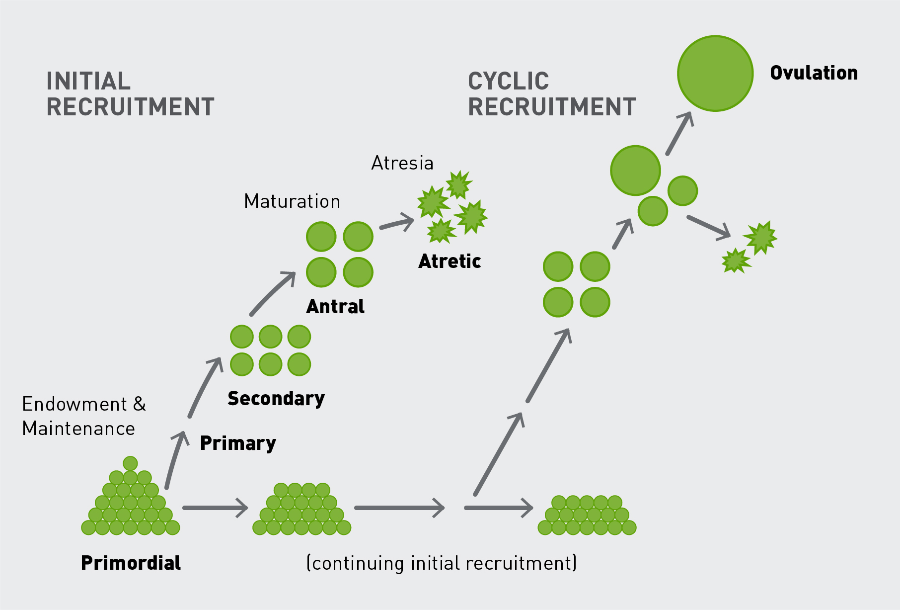
Figure 3: The follicular wave of the cow
Hormones
The oestrous cycle is controlled by a series of hormonal interactions. A deep understanding of these hormonal interactions and how they control the ovarian function enables synchronisation protocols to be developed and manipulated. Below is a brief description to help you understand the basics of each hormone in the oestrous cycle:
-
Gonadotrophin Releasing Hormone (GnRH): Released from the hypothalamus in the brain, it stimulates the release of FSH and LH. Pharmaceutical preparations of GnRH tend to have more LH activity.
-
Follicle Stimulating Hormone (FSH): A surge in circulating FSH acts on the ovaries to stimulate the initiation of a new follicular wave. Transient release of FSH ensures follicular growth assisting in follicle development, maturation prior to ovulation.
-
Luteinising Hormone (LH): A surge in LH induces ovulation in follicles that tend to be dominant or greater than 10 mm in diameter. Transient release supports follicular growth.
-
Oestradiol (E2): Produced by the dominant follicle. When E2 levels are maximised it promotes a surge in LH circulation. E2 is responsible to causing oestrus behaviour.
-
Progesterone (P4): produced by the CL, and prevents oestrus behaviour. Pharmaceutical preparations of P4 are used to mimic the luteal phase of the oestrous cycle. Hence can be used to stimulate anoestrous/prepubertal cattle to cycle. Whilst P4 levels are maximal, FSH and LH release is suppressed. P4 is also responsible for maintaining pregnancy.
-
Prostaglandin F2α (PGF2α): Also known as PG. Secreted by the uterus with the primary action of causing luteolysis or regression of the CL.
A detailed description of the hormonal interactions and how they influence the ovarian function and oestrous cycle can be found in vet resources.
Anatomy
Figure 4, outlines the basic anatomy of the bovine reproductive tract. The reproductive tract of the cow/heifer consists of the vagina, the cervix, two uterine horns, two oviducts, and two ovaries. The function of each part of the reproductive tract relevant to artificial reproduction technologies are as follows:
-
Vagina: pathway to the uterus, where insemination or embryo transfer pipettes are traversed through to the cervix and uterus. Once a Cue-Mate® has been placed into the cow/heifer, it will remain in the vagina for the duration of treatment.
-
Cervix: provides a barrier between the uterus and the vagina. During oestrus the cervix will produce a mucus that assists in sperm transport. During artificial insemination or embryo transfer, the pipette is passed through the cervix to reach the uterus.
-
Uterus: assists in sperm transport to the oviduct. The uterus is responsible for housing the foetus throughout gestation.
-
Oviduct: the site in which fertilisation occurs. Important changes to sperm function occur here transforming the sperm to become capable of fertilisation. The oviduct may store the sperm for a short period of time, whilst waiting for ovulation to occur.
-
Infundibulum: responsible to ‘catching’ the oocyte after ovulation and transporting it to the oviduct.
-
Ovary: houses the oocyte (egg) prior to ovulation. It is responsible for ovulation and subsequent development of the corpus luteum (CL). It also plays an important role in hormone production.
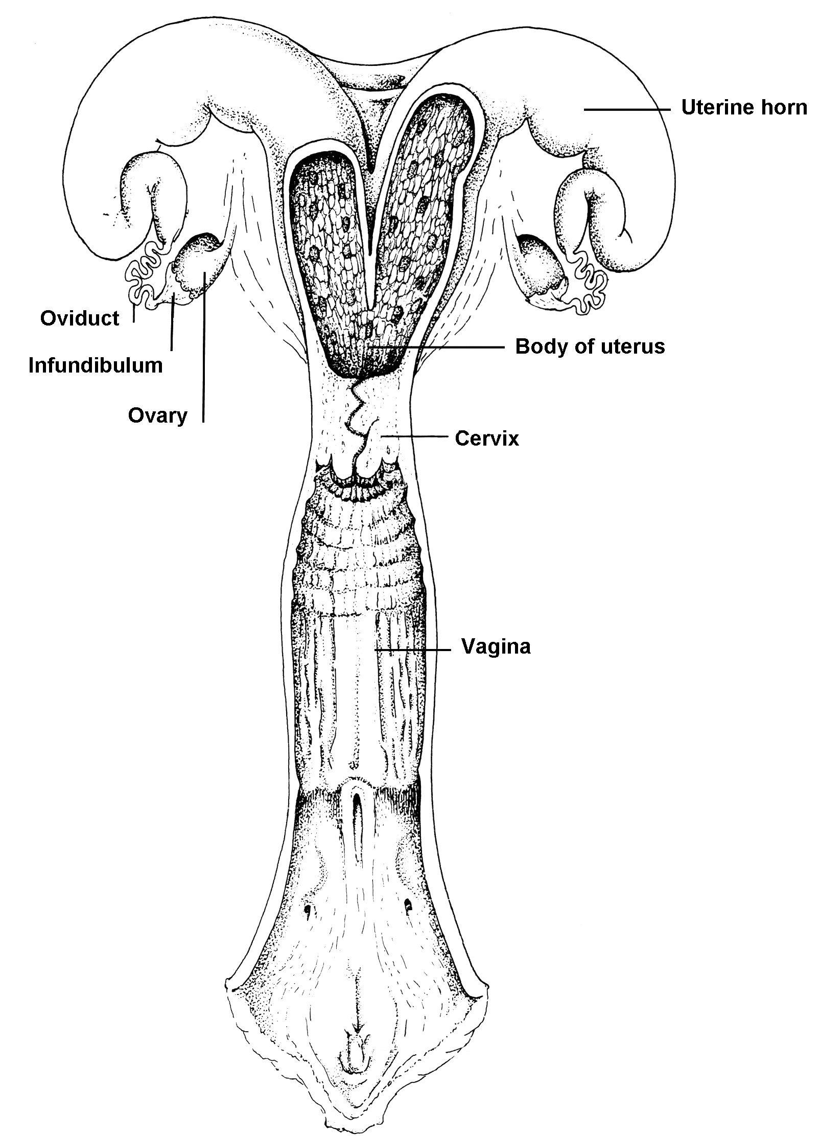
Figure 4: Basic anatomy of the bovine reproductive tract